


Describe the anatomical structure of the dicot root and compare it with the monocot root.
1 Answer
The Dicot Root:
- Epiblema or epidermis or piliferous layer: It is the outermost uni-layered with several unicellular root hairs. It consists of thin-walled, compactly arranged living parenchymatous cells. Epiblema of a root differs from the epidermis of the stem in being devoid of distinct cuticle and stomata. Due to the presence of unicellular root hairs, it also helps in the absorption of water and minerals from the soil.
- Cortex: It lies below the epiblema. The cortex is made up of many layers (6 to 8) of thin-walled oval, rounded, or angular parenchyma cells with intercellular spaces for the exchange of gases. The cells of the cortex store food. They also conduct water from the epiblema to the inner tissues.
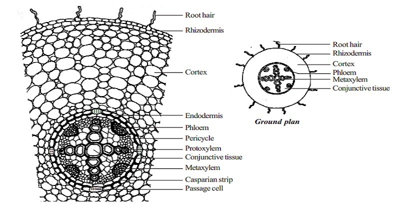
- Endodermis: It is the innermost layer of the cortex and covers the stele. It consists of compactly arranged barrel-shaped parenchyma cells without intercellular spaces. Most of the cells are characterized by the pressure of special thickening of suberin and lignin on their radial and tangential walls and called Casparian strips. Some endodermal cell near protoxylem has no casparion strips and is called passage cells or transfusion cells. These cells allow radial diffusion of water and minerals through the endodermis.
- Pericycle: The layer next to the endodermis is known as the pericycle. The cells of the pericycle are thin-walled and parenchymateous without intercellular spaces. Pericycle helps in the formation of the lateral meristem. At the time of secondary growth, it produces secondary cambium or phellogens.
- Vascular bundle: Vascular bundles are two to eight in number, radial, and arranged in a ring. Xylem and phloem bundles are separated from each other by parenchymatous cells called conjunctive or complementary tissues. Xylem is exarch and consists of tracheids, vessels, parenchyma, and fibers. Phloem consists of sieve tubes, companion cells, and phloem parenchyma. Usually, phloem fibers are absent or reduced.
- Pith: It is feebly developed and centrally located. It consists of thin-walled, polygonal parenchymatous cells without intercellular spaces. In dicot roots, it may be reduced or absent. It helps in the storage of food material.
Monocot Root:
- Epidermis or Epiblems: The epidermis or outermost layer is commonly known as the epiblema or piliferous layer. It is uni-seriater and composed of compact tubular cells having no intercellular spaces among them. The outer regions of epiblema contain unicellular root hairs which help in the absorption of water and minerals from the soil.
- Cortex: Immediately beneath the epidermis a massive cortex lies consisting of thin-walled parenchyma cells having intercellular spaces. Usually, in an old root, a few layers of cortex immediately beneath the epiblema undergo suberisation and give rise to single or multilayered zones of exodermis. This is a protective layer that protects internal tissue outer agencies.
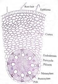
- Endodermis: It is the innermost layer of the cortex and covers the stele. It consists of compactly arranged barrel-shaped parenchyma cells without intercellular spaces. Most of the cells are characterized by the presence of special thickening of suberin and lignin on their radial and tangential walls and are called Casparian strips. Some endodermal cell near protoxylem has no Casparian strips and called passage cells or transfusion cells. These cells allow radial diffusion of water and minerals through the endodermis.
- Pericycle: It lies just below the endodermis and is composed of thin-walled parenchymatous cells without intercellular space (sometimes patches of sclekenchymatous cells are also found). Several lateral roots arise from this layer.
- Vascular bundle: Vascular bundle is radial, arranged in a ring. Xylem and phloem are present in a separate bundle and are numerous and referred to as polyarch. The phloem bundle consists of sieve tubes, companion cells, and phloem parenchyma. Xylem bundle consists of protoxylem which lies towards the periphery and met xylem towards the connected by parenchymatous tissue called conjunctive tissues.
- Pith: Pith is a large, well-developed portion of monocot roots. It occupies the central portion and made from thin-walled parenchymatous tissues with intercellular spaces. It contains an abundant amount of starch grains.

Topics from Biology
Related Questions